Our results
Take a look at the gallery of treatment results
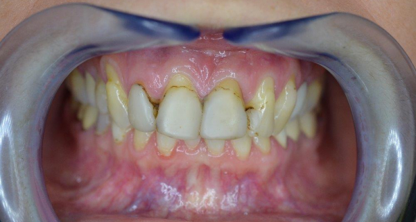
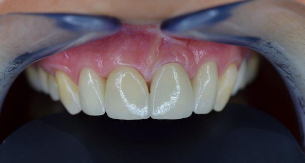
EXPOSED NECKS AND CERAMIC E.MAX CROWNS
Before the procedure
After the procedure
The course of the procedure
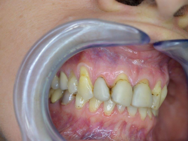
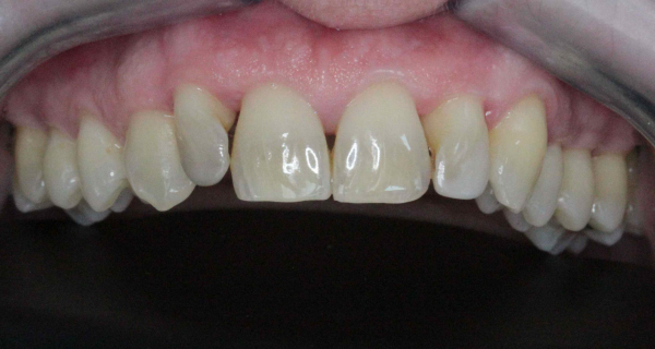
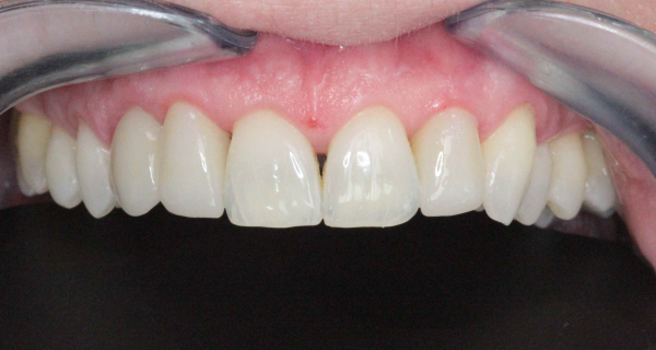
A patient with gum recesses and very unsightly composite facets. We indicated periodontal surgery (by MUDr. Jan Dražan – Dentkomplex, s.r.o.), who returned the gums to their original place and treated the teeth with ceramic e.max crowns. Instead of taking a traditional impression, the prepared teeth were scanned with a TRIOS dental scanner and the ceramic crowns were made by the Keramika Kulovaný, s.r.o. dental laboratory.
Service detailCERAMIC FACETS AND SMILE DESIGN
Before the procedure
.jpg)
After the procedure
.jpg)
The course of the procedure
A patient with white fillings in the anterior section of the denture that have already lost their good looks. Especially visible are pigments at the interface of fillings and teeth. We opted for four e.max ceramic facets. The preparation was scanned with a TRIOS dental scanner. Our Keramika Kulovaný, s.r.o. dental laboratory prepared proposals for the future situation using Insignia Advanced Smile Design to better consider the patient’s wishes. The requirement was to maintain the “butterfly wings”, i.e. a slight “internal” rotation of the large upper incisors. We also used software to find the ideal shape for the small upper incisors. Photographs of the final work were taken less than three weeks after the procedure. We are still waiting for gum regeneration, which is slower.
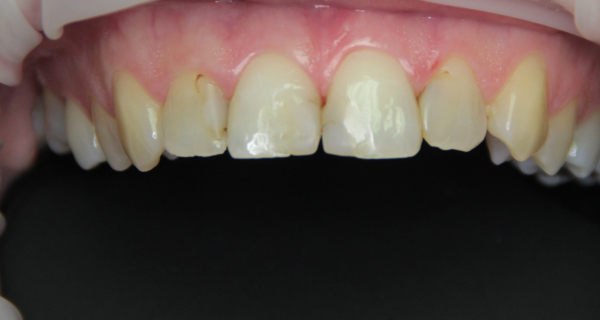
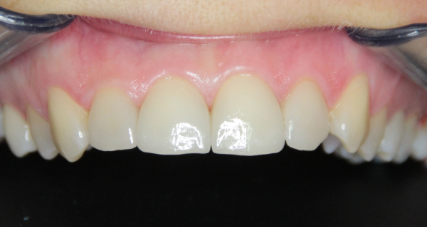
Service detailALL-CERAMIC E.MAX CROWNS OF UPPER INCISORS
Before the procedure
After the procedure
The course of the procedure
Upper incisors with inappropriately modelled white fillings, under which there is also tooth caries. Due to the scope of treatment of the upper incisors, we decided for four e.max all-ceramic crowns. For the result after 1 year, see the gallery.
Service detail4 E.MAX CROWNS AND CROSSBITE OF CANINES ON THE RIGHT SIDE
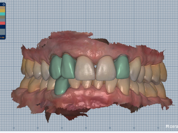
A quick aesthetic transformation without orthodontics? Yes, it is possible. At our clinic Stomatologie Ujec, s.r.o., we recently performed a complete smile reconstruction for a patient who wanted a beautiful and natural smile - without long-term orthodontic treatment. The result? A transformation within a few days, thanks to advanced technologies, precise work and the experience of our team. What the procedure entailed: Teeth whitening with the premium Pure Whitening system, Factoring of 4 all-ceramic e.max crowns on the rotated upper second incisors and canines on the right side, which were in a crossbite. The patient originally considered an orthodontic solution, but due to the time demands and personal preferences, she chose a faster alternative without straightening her teeth with braces. Thanks to detailed digital diagnostics and careful planning, we managed to achieve an exceptionally natural result that met both aesthetic and functional requirements. The treatment was performed by: MUDr. Alexandr Ujec – specialist in smile reconstruction and working with CAD/CAM technologies Technologies used: Trios 3Shape intraoral scanner – precise digital impressions without unpleasant impression material, Ceramill Mind CAD software – 3D design of custom crowns, Digital Smile Design – planning a smile in advance based on the patient’s wishes and facial morphology. Result? The patient left with a completely new, fresh and natural smile – without the need to wear braces, without pain, and most importantly in record time.
Service detailDEEP FRACTURE OF THE UPPER SMALL INCISOR
Before the procedure
.jpg)
After the procedure
.jpg)
The course of the procedure
The patient arrived soon after an accident at work, which resulted in the upper small incisor on the right side being broken. The fracture reached the level of the bone from the palate and, of course, also reached the cavity with the dental pulp (nerve). The root canal was treated, an aesthetic extension was prepared and the tooth was prepared for an e.max all-ceramic crown. The whole treatment was performed under a microscope with 25x-40x magnification and the impression was transferred to the laboratory using a 3shape TRIOS dental scanner. The result is an exact and aesthetically and biologically compliant all-ceramic e.max crown with no gum irritation.
Service detailZIRCONCERAMIC CROWN AND ROOT CANAL OF THE UPPER LARGE INCISOR
Before the procedure
.jpg)
After the procedure
.jpg)
The course of the procedure
The patient needed treatment of a darkened upper large left incisor with numerous fillings and with a large brown spot by the cutting edge. The original plan was “only” to treat the canal, whiten the tooth and make new fillings. But after removing the fillings and small caries, only the front lamella of the tooth remained; the back had gone all the way down to the gums. Therefore, after completing the treatment of the root canal, we decided to make preparation for a zirconceramic crown. You can see the result in the gallery.
Service detailFACET VS. E.MAX ALL-CERAMIC CROWN
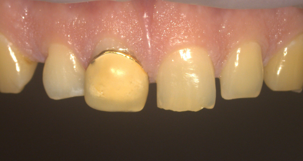
Before the procedure
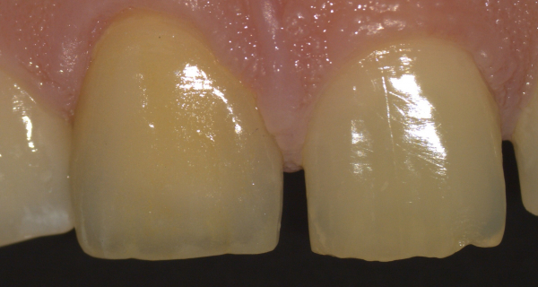
After the procedure
The course of the procedure
In his adolescence the patient suffered an injury to the right upper incisor, which had been treated with a gold-resin facet crown (state of the art at the time). The gold alloy allowed casting into thin layers and allowed minimal preparation, saving the dental pulp (nerve). Nowadays, with the help of minimally invasive procedures, adhesive fixation methods, using a microscope and other technologies, we do not have to be afraid of making either a larger photocomposite extension or an all-ceramic crown. After years of gold resin work, we made the patient’s smile look natural again. An E.max all-ceramic crown was made using a Leica dental microscope and scanned for the laboratory with a Trios scanner.
Service detailALL-CERAMIC E.MAX OVERLAY
Before the procedure
.jpg)
After the procedure
.jpg)
The course of the procedure
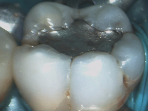
The patient came with a partial fracture of a tooth in a large amalgam filling, which we temporarily treated. During the subsequent preparation, we chose how to “hold” the ceramic material. The choice was an overlay, which will have greater retention than an e.max crown. Preparation for the e.max crown would have been more radical and weakened the tooth even more. See the result in the gallery.
Service detailNAVIGATED ENDODONTICS OF THE UPPER SMALL INCISOR WITH OBSTRUCTED ROOT CANAL
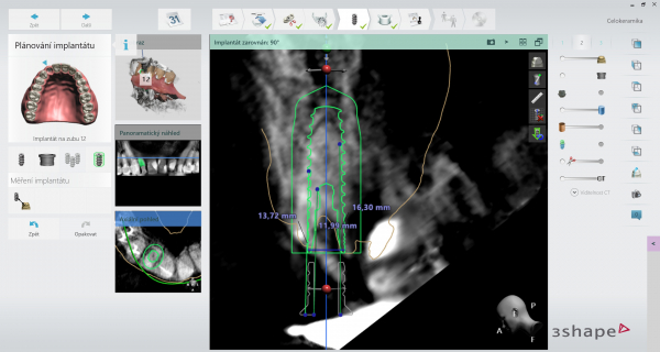
Before the procedure
After the procedure
.jpg)
The course of the procedure
The patient came with a loose filling and part of the upper small incisor. Eight years ago, this filling was made due to a large area of caries. Endodontics (root canal treatment) and insertion of a glass-composite pin into the filled canal were indicated to retain the crown part of the tooth. However, the previous endodontics from eight years before was not successful due to considerable obliteration (calcification) of the root canal. Now, after part of the filling and tooth broke off, we used a dental scanner with a dental CT. Thanks to this data, a 3D printed template for a computer-guided root canal treatment was created. The result is a hermetic root filling of the entire root canal and the introduction of an extension pin for the fitting of an all-ceramic crown.
Service detailBROKEN TOOL REMOVAL PEN (BROKEN TOOL REMOVAL PEN)
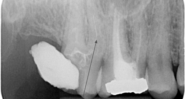
Before the procedure
After the procedure
The course of the procedure
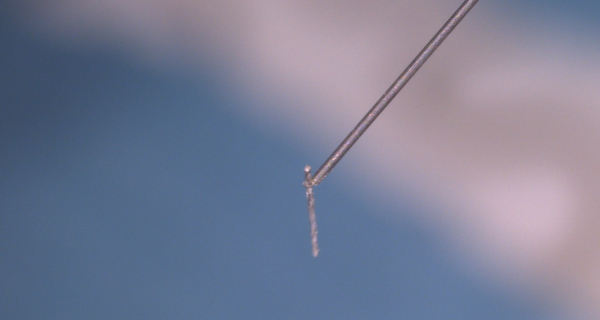
The patient came to the entrance examination, during which a broken root instrument was found in the second upper stool from the previous nurse. Once freed, this fragment was removed using a BTR-PEN with a microloop, which is threaded under a microscope onto the kinked root instrument in the root canal. After the extraction, the treatment of the root of the tooth was completed.
Service detailREMOVING A BROKEN TOOL FROM THE UPPER MOLAR
Before the procedure
 (1).jpg)
After the procedure
 (1).jpg)
The course of the procedure
The patient came from her doctor needing the removal of a broken-off tool under a microscope. This is an infrequent complication of root canal treatment that needs to be addressed, as it would reduce the likelihood of long-term success of this treatment. After some time, the tool was removed using a special ultrasound and then the tooth with four root canals was treated, filled and finished with a photocomposite material (white filling). The last image shows that small infectious sites around the tooth have healed and the prognosis for the whole tooth is now much better than at the beginning.
Service detailREMOVAL OF A BROKEN ROOT TOOL FROM THE TOOTH CANAL AND SUBSEQUENT PROSTHETIC TREATMENT
Before the procedure
After the procedure
The course of the procedure
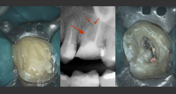
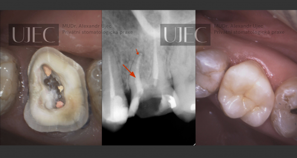
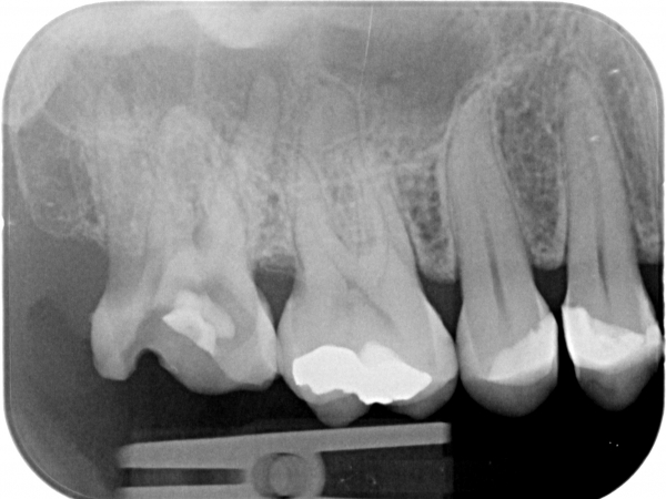
In another dentist’s office, a tool was bent in the root canal of the upper left first molar and an inflammation formed above this root. The treatment with the help of a dental microscope was demanding, but successful. The tool was removed and after hermetically filling the canals, we finished the tooth with an e.max all-ceramic crown. From left: tooth with temporary filling; X-ray image with location of the broken root canal tool (large arrow) and inflammation (small arrow); preparation of the access to the canal concerned From the left: a filled canal (upper edge of the tooth) and preparation for the impression for the e.max crown; X-ray image showing hermetic filling (arrows); fitted e.max ceramic crown
Service detailREMOVING A BROKEN TOOL FROM THE ROOT CANAL
Before the procedure
After the procedure
The course of the procedure
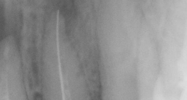
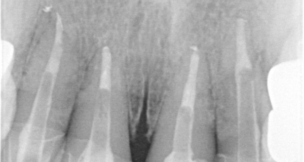
During initial examination, we found from X-ray images that this patient had a foreign object in the root canal of the upper left canine, i.e. a broken root tool. This was an accident during treatment, which has a complicated solution. If this tooth were not treated or this foreign object could not be removed, over time the tooth would have to be extracted and it would have to be replaced, for example, with an implant. You can see the whole procedure in the gallery of the procedure. The condition of the upper left canine before treatment: in the middle of the canal there is a broken instrument from the previous dentist. In addition, the tooth is affected by chronic inflammation around the tip of the root. If the foreign body cannot be removed from the root canal, there is a risk that we will have to extract the tooth and replace it with, for example, an implant.
Service detailDELEGATED PATIENT FOR ROOT CANAL FILLING COMPLICATIONS
Before the procedure
After the procedure
The course of the procedure
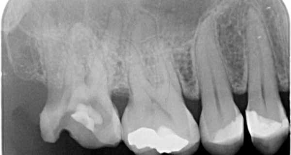
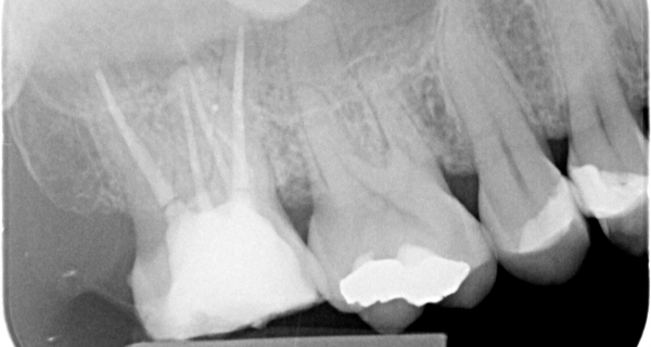
The patient was referred by her dentist for the impossibility of completing the root canal filling. The X-ray image shows an anomalous configuration with five roots. After preparing the tooth, we probed and then treated 5 root canals. From the available literature, this configuration is described for tooth 6 in the lower jaw in approximately 10% probability of occurrence.
Service detailTREATMENT WITH NEW ROOT FILLING AND HEALING OF CHRONIC INFLAMMATION NEAR THE ROOT
Before the procedure
After the procedure
The course of the procedure
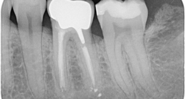
A 28-year-old patient with a completely inappropriate root filling of the lower left first molar with a chronic infectious lesion around the root. Again, treatment of root canals was indicated. We placed a precise metal-ceramic crown on the tooth. After one year, we observed the complete disappearance of the chronic lesion around the root. The tooth with completely unsatisfactory root filling and chronic inflammation around the root (dark areas resembling arches or even circles under the roots of the tooth) Tooth with a new 3D hermetic root filling and sprayed branching of the root canal of the posterior root (on the right side of the image) Control X-ray after 1 year: tooth with a precise metal-ceramic crown and no more inflammation around the root, which healed during this relatively short time
Service detailEXTENSIVE CARIES AND INFLAMED LESION ON THE LOWER FIRST MOLAR WITH SUBSEQUENT TREATMENT
Before the procedure
After the procedure
The course of the procedure
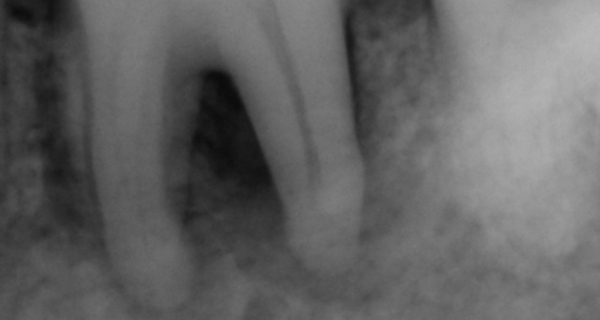
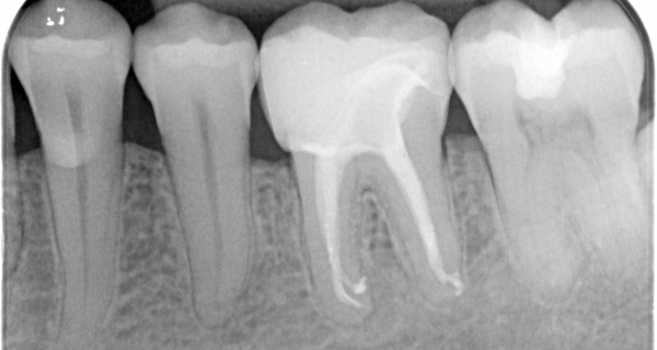
Lower left first molar: tooth with amalgam filling, extensive caries affecting the dental pulp (nerve) and infected root system. There was a large infectious deposit in the surrounding bone at the root of the tooth. We removed the caries, treated the root canals and restored the tooth with an e.max with an all-ceramic crown. After 6 months, remineralisation of the bone, i.e. healing of the inflammatory deposit, can be seen. The ceramic e.max crown meets all requirements for both function and aesthetics.
Service detailCERAMIC E.MAX CROWNS, AESTHETIC ROOT EXTENSION AND COMPOSITE FILLINGS
Before the procedure
.jpg)
After the procedure
.jpg)
The course of the procedure
The patient was, of course, dissatisfied with the “aesthetics” of her smile. She was literally ashamed to smile. What kind of life is it when you are afraid to smile...? The therapeutic plan indicated a revision of the root canal of the large upper right incisor, followed by an aesthetic root extension using a glass-composite pin. It was also necessary to replace the fillings of both small incisors. (There are other treatments planned, but they are not so urgent.) The result was photographed 4 days after the fitting of e.max crowns. Judge for yourself.
Service detailRECONSTRUCTION OF THE ENTIRE UPPER JAW – ZIRCONCERAMIC BRIDGE, E.MAX CROWNS AND PROSTHETIC TREATMENT OF IMPLANTS
Before the procedure
After the procedure
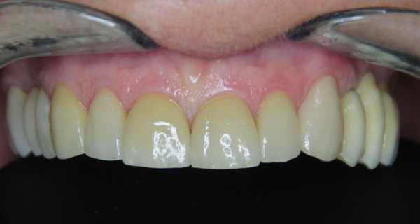
The course of the procedure
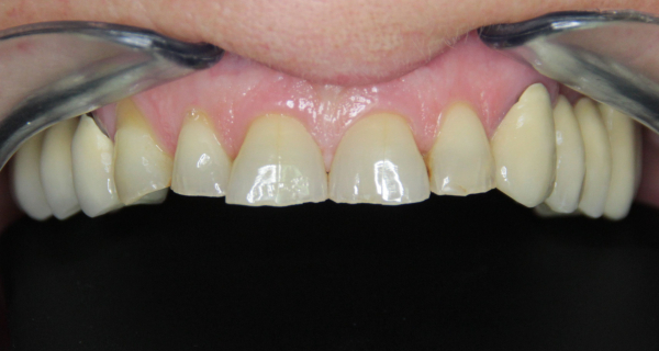
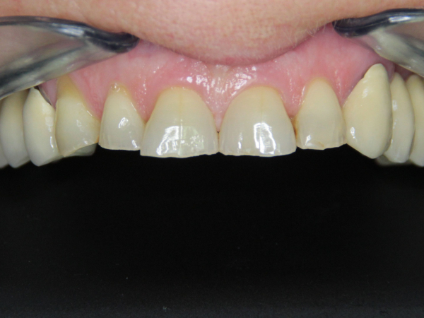
In the patient, we indicated preparation for the e.max crown in the anterior section of the upper jaw, as the incisors and right canine were heavily abraded (shortened by grinding by articulation of the lower metal-ceramic bridge (which we also replaced) and by gradually sliding the metal-ceramic bridge against the upper jaw). Furthermore, in the back section of the right side (left side in the pictures) we made a new zirconceramic bridge and on the left side (right side in the pictures) it was necessary to insert two implants (after extraction of the first molar; extracted by MUDr. Jan Dražan), on which a zirconceramic bridge was also fitted after integration into the bone tissue. All procedures were performed using a Leica dental microscope and scanned using a Trios scanner. The result is well worth it.
Service detailIMPLANTATION AND PROSTHETIC TREATMENT OF SHORTENED DENTAL ARCH
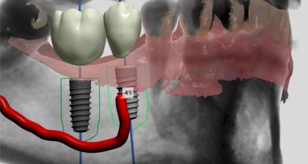
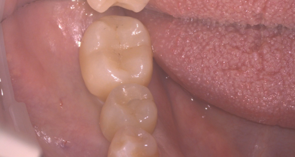
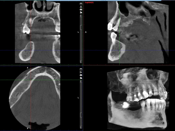
The patient was scheduled for two implants in the missing place 6 and 5 on the lower right. CT navigation was used to accurately introduce and achieve parallelism of the preparation. A template determining the direction and depth of the preparation was designed by combining the digital impression made by the dental scanner and CT. After the implants had healed, the prosthetic treatment with all-ceramic crowns was completed.
Service detailTREATMENT OF PERIODONTITIS (PERIODONTOSIS), AESTHETIC FILLING, ALL-CERAMIC FACETS AND CROWNS
Before the procedure
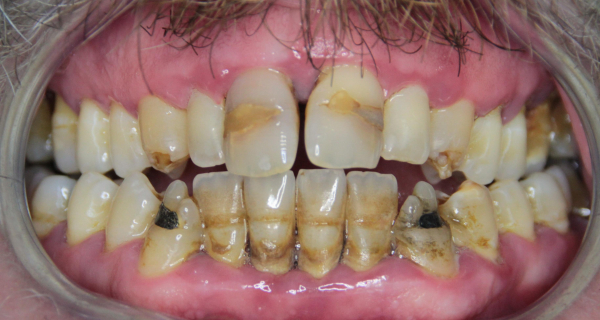
After the procedure
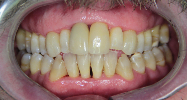
The course of the procedure
Patient with ongoing periodontitis, inappropriate “white” fillings, enamel deformities after previous applications of tetracycline antibiotics, amalgam fillings (formerly this indication was state of the art) of the lower canines. In cooperation with MUDr. Jan Dražan, we treated the patient and ended the ongoing periodontitis, followed by the production of photocomposite white fillings, e.max all-ceramic facets and crowns. The result is excellent aesthetics, where the gums are receded, but without ongoing inflammation. Everything was done under a Leica dental microscope.
Service detailCOMBINED TEETH WHITENING - PUREWHITENING
Before the procedure
.jpg)
After the procedure
.jpg)
The course of the procedure
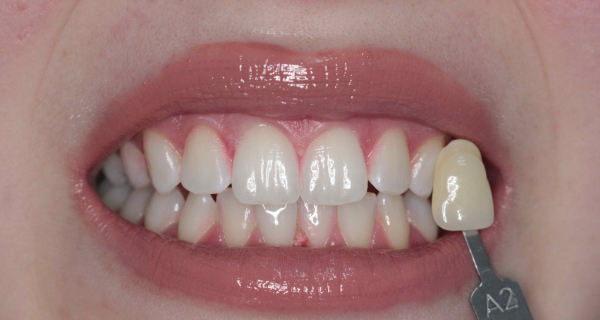
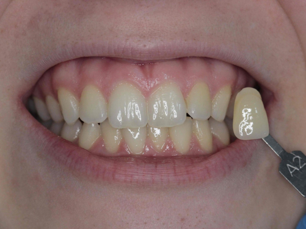
The patient with tooth shades B3/C2 underwent combined whitening with Purewhitening, which consists of home and in-office phases. The result is shade B1 for all teeth. The white diet (elimination of coloured foods, coffee and smoking) was not followed at all. The patient enjoys a lot of coffee and cigarettes, especially in the summer months, when the whitening took place. See the result in the gallery.
Service detailHOME TEETH WHITENING - PUREWHITENING
Before the procedure
.jpg)
After the procedure
.jpg)
The course of the procedure
The patient chose Purewhitening home. The aim was a slight lightening of the tooth colour, which can be seen in the picture after the completion of home whitening compared to the original shade A3.
Service detailBLANCONE CLICK WHITENING
BlancOne Click - 15-minute whitening performed after dental hygiene. Boosted with a blue spectrum LED whitening lamp. Without sensitivity and limitations. Instant result.
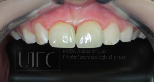
Service detailINJURY OF A 9-YEAR-OLD – BROKEN CROWNS OF THE UPPER INCISORS DOWN TO THE DENTAL PULP (NERVE)
Before the procedure
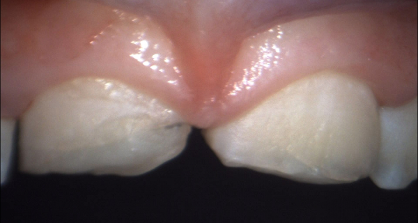
After the procedure
The course of the procedure
A boy of 9 years – an injury on skates, a fall on ice – a complicated fracture of the crowns of both permanent incisors. Both teeth had two point perforations in the dental pulp (nerve). In addition, the root development of both incisors was not yet complete. The treatment was started 50 minutes after the injury and in the first phase involved “cementing” the perforations (holes) in the dental pulp in order to keep the teeth (dental pulp) alive so that they could complete the root development. Only after 5 months did we make all-ceramic e.max crowns. The impression was digitally transmitted by a Trios scanner to the dental laboratory of Keramika Kulovaný, s.r.o. The teeth are still vital. In the future, it is expected that the crowns will be redesigned according to the new conditions around the second decade of the life of the young patient.
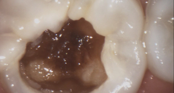
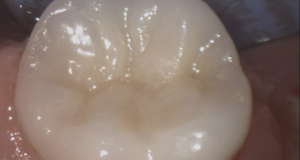
Service detailCHILD AGED 10 TREATMENT OF LOWER FIRST MOLAR ROOT CANALS AND E.MAX CERAMIC CROWN
Before the procedure
After the procedure
The course of the procedure
A boy of 10 years of age came for initial examination, where we diagnosed caries in the lower right first molar extending into the dental pulp (“nerve”). One of the treatment options is tooth extraction and subsequent orthodontic treatment. It is a long-term treatment. A decision was made with the parents and the boy to save the tooth. The prognosis of treatment will be long-term, although it may not be lifelong. It is possible that in late adulthood it will be necessary to extract the tooth and insert an implant or autotransplant one of the third molars (wisdom teeth). The production of the e.max ceramic crown is performed by our dental laboratory Keramika Kulovaný, s.r.o.
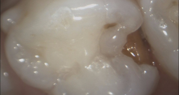
Service detailDECIDUOUS TEETH FILLING – CASE HISTORY
Before the procedure
After the procedure
The course of the procedure
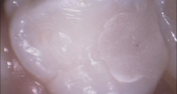
Deciduous teeth are a sort of small version of the permanent tooth. However, they differ in some aspects, such as the proximity of the dental pulp (nerve) to the tooth surface. This requires increased treatment requirements. We work exclusively with magnifying optics to achieve excellent results in such a small space. Take a look at the gallery.
Service detailAESTHETIC PHOTOCOMPOSITE FILLINGS
Before the procedure
After the procedure
The course of the procedure
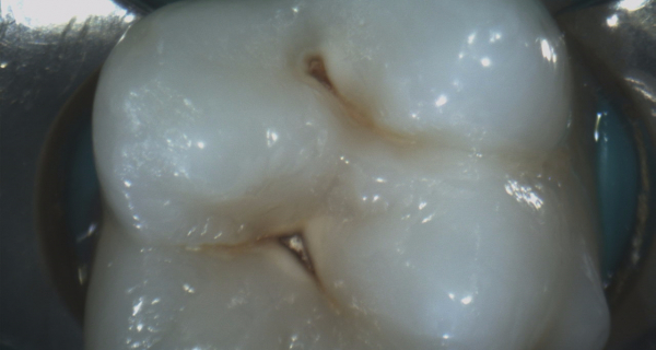
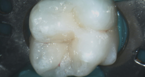
We ensure that the filling must not be visible. We perform preparation and modelling under a Leica dental microscope and in latex insulation from the oral cavity. We ensure complete removal of tooth caries. We do not compromise that would worsen the durability of the treatment.
Service detailDENTAL HYGIENE – MOTIVATED PATIENT
Before the procedure
After the procedure
The course of the procedure
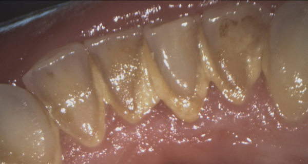
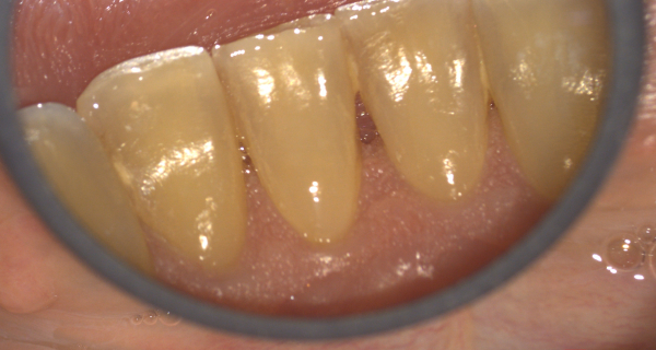
The patient (44 years old) visits us regularly for preventive check-ups and dental hygiene. During the two-year period (between 2017-2019), we achieved motivation and improved teeth-cleaning techniques with the patient during four dental hygiene visits. He uses a Philips Sonicare electric toothbrush, flosspick, interdental brushes and a solo brush to finish cleaning. Measured by a stopwatch – evening dental hygiene within 4.5 minutes See for yourself in the gallery, where we display a series of images from preventive check-ups before visiting a dental hygienist.
Service detail
.jpg)

.jpg)
.jpg)

.jpg)
.jpg)

 (1).jpg)


.jpg)

.jpg)
.jpg)



.jpg)
